Tag Archives: Biology

Antibiotic resistance: a challenge to rival climate change

Dr Clare Sansom, Senior Associate Lecturer in Birkbeck’s Department of Biological Sciences reports on a recent, international capacity-building workshop to tackle antimicrobial resistance and accelerate new antibiotic discovery, sponsored by the global challenges research fund (GCRF). 

Credit: Mr Harish Patel

Antibiotic resistance is one of the most serious threats to global health: some commentators have even rated it as important as climate change. It is, however, one that the research community – particularly in the academic and not-for-profit sectors – is finally investing serious resources in tackling. Research into novel antibiotic targets and the compounds that can interact with them is burgeoning throughout the world. At Birkbeck, research in the ISMB-Microbiology Research Unit, headed by Professor Sanjib Bhakta focuses on tackling antibiotic resistance in priority bacterial pathogens, the causative agent of a number of global infectious diseases.

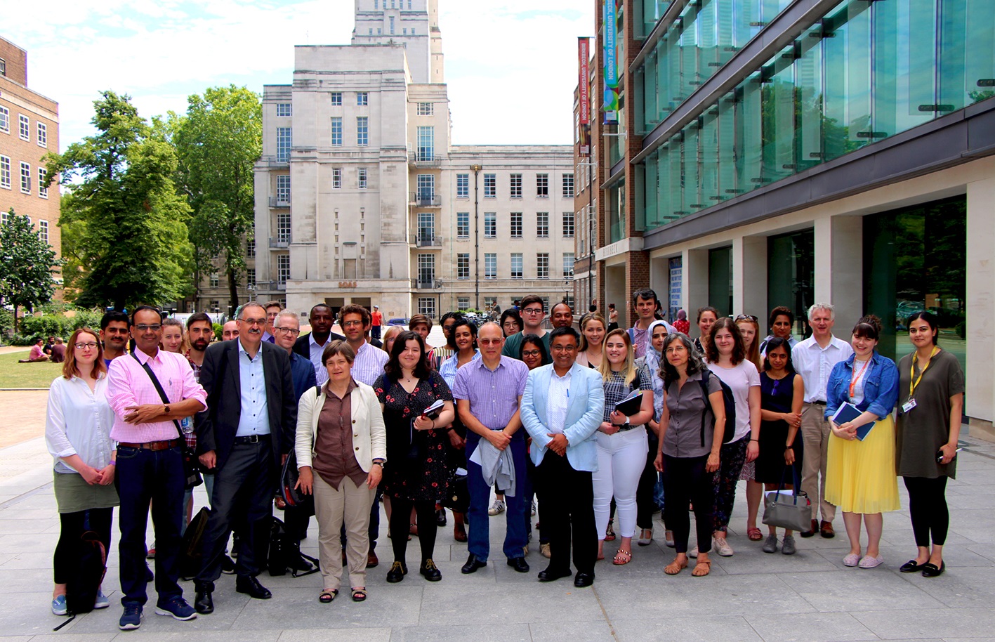
In July 2019 Prof Bhakta invited his collaborators in the UK and overseas to a workshop at Birkbeck to discuss ways of tackling drug resistance. This was funded through the UK’s Grand Challenges Research Fund (GCRF), an initiative to promote the welfare of developing countries through international research. Delegates were welcomed by Birkbeck’s Pro-Vice-Master for internationalism, Professor Kevin Ibeh, and then heard brief presentations from Dr Sarah Lee on the place of the GCRF in Birkbeck’s research portfolio and Dr Ana Antunes-Martins on the mission of the neighbouring London International Development Centre. This combines the resources of seven University of London institutions in the Bloomsbury area, including Birkbeck, to support interdisciplinary research, capacity building and public engagement for international development.

In an intense scientific session, the first researcher to speak was Professor Nicholas Keep, Executive Dean of the School of Science at Birkbeck and a structural biologist. He presented some structures of Mycobacterium tuberculosis proteins known or believed to be novel prospective therapeutic targets that had been solved at Birkbeck or UCL using the three main techniques of structural biology: X-ray crystallography, nuclear magnetic resonance (NMR) and electron microscopy (EM). These included a small enzyme called resuscitation-promoting factor, which is necessary for the bacteria to emerge from their dormant state, and a rather larger one that synthesises an important component of the cell wall. This work is enabled by excellent local facilities including a new Titan Krios microscope in Professor Helen Saibil’s EM lab, and even more powerful national and international ones.

Beta-lactamase enzymes, which inactivate drugs in the penicillin family, are one of the most common antibiotic resistance mechanisms, but they are not as well understood in mycobacteria as they are in some other human pathogens. Prof Anindya S. Ghosh from the Indian Institute of technology in Kharagpur – incidentally, the speaker who had travelled the furthest – described his group’s work in collaboration with Prof Bhakta and Prof Tabor’s lab (funded by a Newton-Bhabha international fellowship to Sarmistha Biswal at Birkbeck this year) in designing inhibitors for beta-lactamases and other proteins that interact with penicillins and prevent their antibiotic action. He set out several more opportunities for collaborative research, including finding molecules that prevent the formation of drug-resistant microbial biofilms.

The next two speakers came from continental Europe, and both described novel natural sources of potential anti-infective drugs. Prof Franz Bucar from the University of Graz, Austria focused on drugs from plant and fungal sources, including an intriguingly named flavonoid, skullcapflavone II (derived from the poisonous skullcap mushroom). This and other recently discovered natural products were also highlighted on a poster by Prof Bucar’s doctoral student, Julie Solnier.  Prof Ester Boix from Universitat Autònoma de Barcelona in Spain described how the antimicrobial peptides that we synthesise as a defence against bacteria might be harnessed as drugs. Her group has used the HT-SPOTi assay developed in Prof Bhakta’s group at Birkbeck to screen human ribonuclease peptides against macrophages infected with Mycobacterium tuberculosis.

Dr Jody Phelan and Prof Taane Clark from the London School of Hygiene and Tropical Medicine asked – and answered – the question ‘What can the M. tuberculosis genome tell us about drug resistance in TB?” This bacterial genome contains over 4 million base pairs of DNA and about 4,000 genes, compared to the 3.3 billion base pairs and 20,000 genes of the human genome. Resistance to any of the over 10 drugs currently used to treat the disease arises largely when treatment is irregular or stopped too soon, and the genetic changes responsible for each type of resistance can be identified rapidly using whole genome sequencing. It is now possible to use this in clinical practice to predict which drugs a given strain is most likely to be resistant to, and thence to recommend a personalised course of treatment for an individual patient.

Dr Simon Waddell from the University of Sussex in Brighton described how the RNA molecules transcribed from the M. tuberculosis genome change during the lifecycle and with the environment of the bacterium, and how this analysis, known as transcriptomics or RNA profiling, can both track and predict responses to drug therapy. One new compound, a benzothiazinone discovered through a high-throughput screen, was found to induce transcription from the same set of genes as cell-wall synthesis inhibitors, suggesting that it is likely to act against the bacterium through the same or a similar mechanism.

These two ‘omics talks were followed by two extremely short ones by scientists based in the Department of Chemistry at University College London. Dr Rachael Dickman (from Prof Alethea Tabor’s lab in UCL) is developing potential antibacterial agents based on a complex amino acid, lanthionine. These ‘lantibiotics’ bind to Lipid II, which is formed during cell wall synthesis, and therefore act as inhibitors of that synthesis. Professor Helen Hailes described the antimicrobial properties of a series of isoquinolines that selectively inhibit slow-growing mycobacteria and that may also potentiate the activity of other drugs by preventing their efflux from bacterial cells.

With the last talk, by Prof Matthew Todd of the UCL School of Pharmacy, the workshop moved from pure science to begin to discuss the economics of drug discovery. Prof Todd’s open source drug discovery work, which began with a project on malaria, is completely open: all the data is freely available, all ideas are shared, no results are ‘owned’ by any of the researchers and there will be no patents. It is a timely approach and one that can involve anyone – Prof Todd has recently been awarded a grant by the Royal Society to work with teenagers at Sevenoaks School to develop new antifungals – and one that might, perhaps, help any of the academic groups represented at the workshop turn their novel ideas into drugs that are useful against the killer disease.

The final networking session was accompanied by an engaging talk by me, Dr Clare Sansom, about the important issue of communicating the challenge of antimicrobial resistance to non-scientists. This was illustrated with frightening scenes from fictional accounts of possible post-antibiotic futures and included a quiz that many of the experts present found surprisingly challenging. This workshop was organised in collaboration with the Commonwealth Scholarships Commission, UK and approved by the Royal Society of Biology for continual professional development credit.

Share

Science Week 2017: fungi in heritage buildings

Dr Clare Sanson, Senior Associate Lecturer in Biological Sciences, writes on Sophie Downes’ talk on fungi and conservation in heritage buildings.mushroom-2198010_1920The Department of Biological Sciences’ contributions to Birkbeck Science Week 2017 focused on ‘Microbes in the Real World’. Apart from that over-arching theme, however, the two sessions could hardly have been more different. The Week kicked off with a lecture by PhD candidate Sophie Downes on the interactions between fungi and heritage buildings. As far as I am aware, Sophie is the first Birkbeck student to have given a Science Week lecture; she spoke with confidence and clarity, and held her audience well.

Nicholas Keep, Executive Dean of the School of Science at Birkbeck, introduced Sophie as a graduate of the University of Lincoln who had worked in textile conservation before moving to Birkbeck to study for a doctorate in Jane Nicklin’s mycology lab. She began her lecture by explaining the context of her research: her job had been based in a large Elizabethan house that had problems with pests and condensation, particularly in the show rooms. The need to find out how best to preserve and repair organic material in buildings like this one led directly to her PhD studies.

In the UK we have a huge number of historic buildings, many of which are popular tourist attractions and play an important role in the local economy, particularly in rural areas. A large number of these are maintained by the National Trust or English Heritage, and many are open to the public for the majority of the year. The thousands of visitors drifting through properties will affect the number and types of micro-organisms, particularly fungi, found there. Sophie’s project included a year-long survey, starting in the autumn of 2013, of fungi found in 20 historic buildings in England, Wales and Northern Ireland. These included cottages and wartime tunnels as well as the more usual castles and mansions, so the survey could be expected to provide a snapshot of fungi and fungal damage in a wide range of historic properties in the UK.

When we think of fungi, we tend to think of so-called ‘macro’ fungi: this category includes the mushrooms we eat and poisonous toadstools, but also dry rot. Micro-fungi are harder to spot, but they are at least as pervasive and colonise an enormous range of organic matter, producing spores. For example, they are responsible for the blue colouration often found on stale bread and preserves. Micro-fungi will colonise almost any organic object that they find in their way, which, in the context of a historic building, might include wood, tapestry, leather book bindings and silk wall hangings. Sophie used air sampling and sterile swabs to obtain representative fungal samples from one outdoor and four indoor locations at each building and recorded the position of and features in each room or area selected, with its temperature and relative humidity.

Sophie landed up with a total of 4,000 samples to analyse, which, given her limited time, was too many for wholescale sequencing. She started by separating these according to colour and morphology and then selected representative samples for DNA extraction and ‘barcode screening’, and fewer for DNA sequencing.  A total of 158 different fungal species from 77 genera were identified, with the most abundant genera being Aspergillus, Cladosporium and Penicillium. Some of the organisms found in smaller quantities, including fungal plant pathogens probably from the outside air and bacteria, were shed from visitors’ skin scales. Both the number of colony forming units and the diversity of fungal species recorded increased during the summer months.

Resident fungi can carry a small risk to human visitors to the buildings and perhaps a slightly higher risk to curators, given their higher exposure times. Fortunately, only a small fraction of the fungi identified were ‘nasty’ human pathogens, and all but one of these were classified in the lowest-risk group, Category 2. A larger number were recognised as of potential risk to particularly vulnerable individuals with damaged immune systems, and more still are only hazardous to the external environment.

The temperature, the height of the building, the type of room and amount of furnishings were found to be the most important factors in determining the extent of fungal growth within buildings and if high colony forming units would be observed, and the three most common fungal species in both the air and the swab samples – Penicillium brevicompactum, Cladosporium cladosporioides and Aspergillus versicolor – have frequently been reported in organic material in historical collections worldwide.

Fungi damage textiles and other organic materials by secreting enzymes that break down polymers, forming secondary metabolic products that cause further degradation. This process has important effects on the physical, chemical and mechanical properties of the materials. Sophie described how she had evaluated each of these, starting with the effect of fungal growth on the physical properties of cotton. Cladosporium infestation is known to cotton fibres, causing an unattractive colour change that cannot be removed by cleaning. She incubated new cotton strips with several fungal species and monitored them for 12 weeks using a technique known as colorimetry. Each fungus caused a gradual colour change, with Cladosporium causing by far the darkest stains. She also reconstructed images of fungi colonising woven cotton fibres in 3D with confocal fluorescence scanning microscopy.

Most fungi have long, filamentous structures called hyphae that secrete enzymes at their tips as they grow. These enzymes break down large and small organic molecules into nutrients; it is the breakdown of large molecules – polymers such as collagen, cellulose, fibroin and keratin – that cause chemical damage to heritage materials. Chitin and keratin are among the most complex organic substrates that fungi can digest and require several enzymes to break them down. Nevertheless, the three commonest species of fungi all managed to reduce the protein content of protein-containing fibres significantly, with Penicillium causing particularly serious damage to collagen. Fungal digestion also changed the local structure of protein fibres. And one net result of this chemical degradation is a change in the mechanical properties of the materials; for example, fungal infestation tends to cause silk to become more brittle.

But what are the implications of these results for the conservation of objects in historic buildings? All the test were conducted on modern materials, and aged ones, which are already worn, are bound to be more vulnerable. Sophie ended a fascinating talk by suggesting that this research will help to inform conservation protocols for the handling, treatment and risk factors involved with fungal contamination of historic collections.

Share

Microtubules and Microscopes: Exploring the Cytoskeleton

This post was contributed by Clare Sansom, Senior Associate Lecturer at the Department of Biological Sciences

"The binding site for End Binding protein 1 (highlighted in green) on the microtubule lattice at the corner of four tubulin dimers, visualised using cryo-electron microscopy" (Credit Cell by Maurer et al (2012))

“The binding site for End Binding protein 1 (highlighted in green) on the microtubule lattice at the corner of four tubulin dimers, visualised using cryo-electron microscopy” (Credit Cell by Maurer et al (2012))

Electron microscopist Carolyn Moores, the most recently appointed professor in the Department of Biological Sciences at Birkbeck, gave her inaugural lecture at the college on June 1.

Moores arrived at Birkbeck in 2004 to start her research group and has risen rapidly and steadily up the academic ladder ever since. Introducing the lecture, the Master of Birkbeck, David Latchman, explained that Moores’ CV stood out in every way; she was clearly as gifted a teacher and administrator as she was a researcher. Furthermore, as she has won several awards for science communication, he predicted that the audience would be in for a treat. We were not disappointed.

Educational journey

Moores began her lecture by saying that she would talk about three different things: her own career development; her group’s research into the structure and function of microtubules; and the advancement of women in science, a cause that is close to her heart.

She remembered that she had wanted to work as a scientist as soon as she knew what a laboratory was, and she started young, as an intern in a research lab at Middlesex Hospital while still in the sixth form. School was followed by a BSc in Biochemistry at Oxford and a PhD in John Kendrick-Jones’ lab at the world-famous Laboratory for Molecular Biology (LMB) in Cambridge. She then moved to work as a post-doc with Ron Milligan at the Scripps Research Institute in La Jolla, California, USA, and it was there that she began her studies on microtubules.

Coming to Birkbeck

The award of a David Phillips research fellowship in 2004 gave her the opportunity to return to the UK as an independent researcher. She explained that there were three reasons – or more accurately three people – that led her to choose to come to Birkbeck. Working in electron microscopy, she was inspired by the work of Helen Saibil, one of the UK’s principal exponents of that technique; she had known Nicholas Keep, then a lecturer in Biological Sciences, as a friend since her time at the LMB; and she knew that she would value the interdisciplinary working environment of the Institute for Structural Molecular Biology under the ‘inspired’ leadership of Gabriel Waksman.

Research into microtubules

Moores then moved on to discuss the main topic of her group’s research: the three-dimensional structure, function and role in disease of tiny cylindrical structures known as microtubules. These are one of the building blocks of the cytoskeleton, which forms a framework for our cells in the same way that our skeletons form a framework for our bodies. They are about 25nm in diameter, which puts them firmly into the ‘nano-scale’ of biology that is easily studied using electron microscopy.

There is a cytoskeleton in every living cell, and it, and the microtubules that form it, are involved in many important cellular processes including shape definition, movement and cell division. Diseases as diverse as cancer, epilepsy, neurodegeneration and kidney disease have been linked to microtubule defects. Understanding their fundamental structure and function, as Moores’ group aims to, should help in understanding these disease processes and perhaps also in developing effective treatments.

Microtubules are built up from many copies of a small protein called tubulin, which, in turn, is a dimer of two similar proteins called alpha and beta tubulin. These tubulin dimers make contacts with each other both head-to-tail and side-to-side to create the cylindrical microtubule wall, fuelled by energy derived from the molecule GTP. Each tubulin unit has a definite “top” and a “bottom” and, as the units are oriented in parallel, so has the complete microtubule.

Microtubules are dynamic structures; they continue to grow by the addition of tubulin units to one end as long as GTP is available, and then begin to unravel and shrink. This dynamism, which allows them to respond to the changing needs of the cell, is essential for their function in healthy cells. In particular, microtubules organise chromosome structures during cell division and are therefore necessary for cell proliferation. As cancer is a disease of uncontrolled cell proliferation, it is possible to imagine that a molecule that could specifically block microtubule growth and assembly in the nucleus might be useful as an anti-cancer drug.

Moores and her group are aiming to understand the process of microtubule growth at as high resolution as possible, using electron microscopy. Unfortunately, however, the most detailed images can only be obtained if the specimen is at very low temperatures (in so-called cryo-elecron microscopy) and using this means that the dynamics of the specimens must be “frozen” into a still image. While it is now possible to see the individual tubulin subunits in the static microtubule images, many details of their structure can only be inferred from computational analysis.

Understanding growth

Moores went on to describe one project in her lab in a little more detail. This was an investigation of the structure and role of proteins that bind only to growing microtubule ends, falling off when the growth stops. It is possible to obtain low-resolution images of microtubules in which these molecules have been made to fluoresce, so only growing microtubules are tracked.

In order to understand the growth process in detail, the group developed an analogue of the GTP “fuel” molecule which can bind to the tip of a microtubule that is extending but not break down to release its energy, so the microtubule does not in fact grow. This forms a static analogue of a growing microtubule that retains all the characteristics of the dynamic structure but that can be studied at low temperatures.

Images of this structure have shown that the end binding proteins bind at the corner of four of the tubulin units. They have explained a lot of the properties of growing microtubules, but there is still more to learn. A full understanding will need structures that are at even higher resolution, where the positions of individual atoms can be made out. Following many years of technical development, today’s most powerful electron microscopes are now making this possible.

Women in science

In the last section of the lecture, Moores left the topic of research to talk briefly about another of her passions: the promotion of women in science. She explained that although 65% of under-graduates in the biological sciences are now women, the proportion of women drops to 40% at any academic grade and 25% for full professors.

A study cited by the European Molecular Biology Organisation has suggested that the barriers for women scientists to progress are set so high that at the current rate of progress full equality would never be achieved. Birkbeck has signed up to the Athena SWAN Charter, set up to encourage higher education institutions to transform their culture and promote gender equality. She described her work with the Athena SWAN team that has so far resulted in the college gaining a bronze award as being as exciting as, but also as challenging as, her studies of microtubules.

Nicholas Keep, Dean of the Faculty of Science and, as Moores had stated, a personal friend, gave the vote of thanks after the lecture. He paid tribute in particular to her value as a colleague, her administrative skills, and the importance of her contribution to the college’s application for the Athena SWAN award.

Find out more

Share