Tag Archives: Biological Sciences

Bernal Lecture 2020: Fifty Shades of Grey Matter

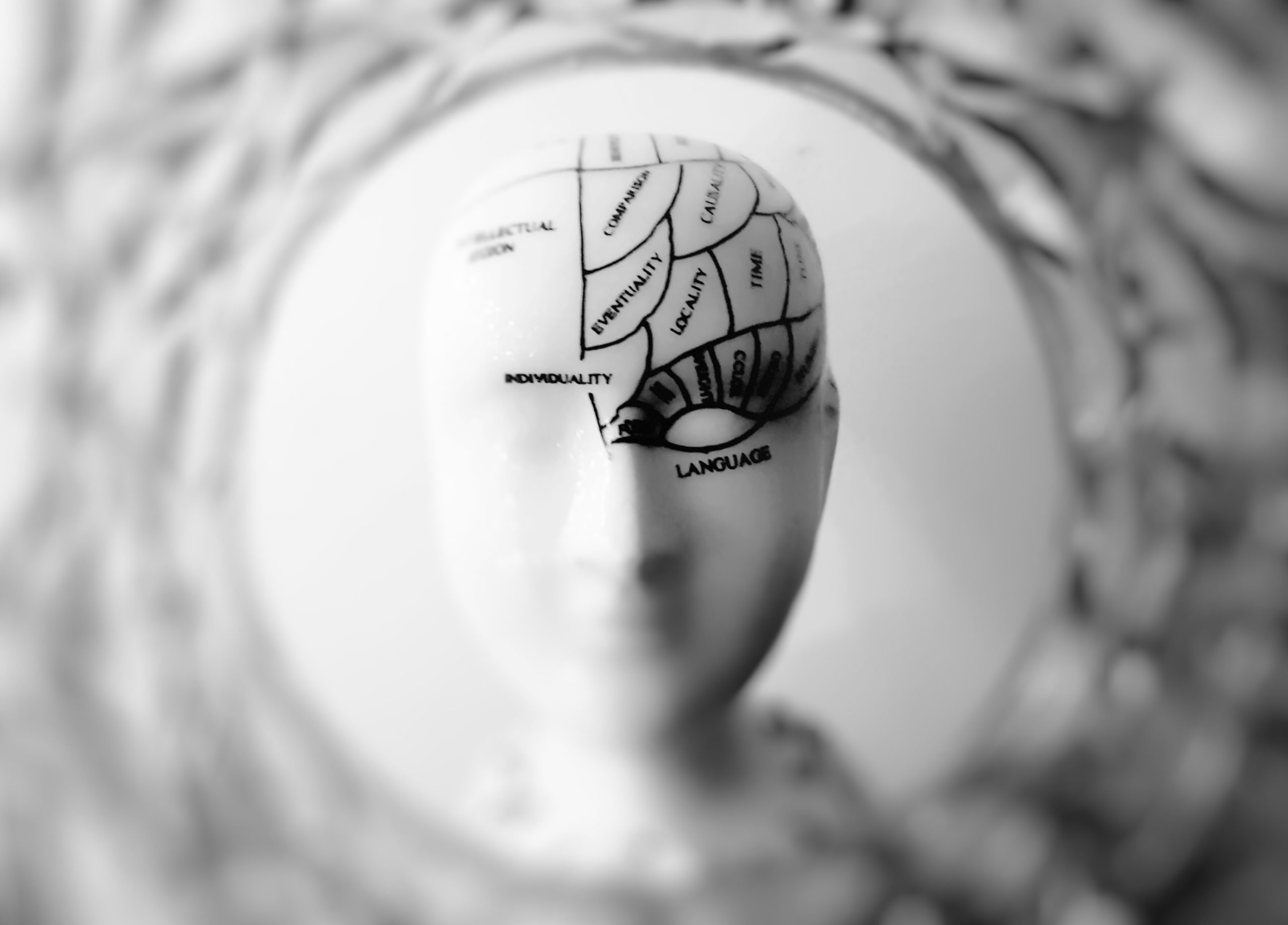
Clare Samson, Senior Associate Lecturer in Birkbeck’s Department of Biological Sciences reports on the annual Bernal lecture that was given by Cordelia Fine, Professor of History and Philosophy of Science on Monday 4 February, that explored whether there really is an essentially male and female brain.

photo of head bust printed

Professor J.D. Bernal is remembered for work on the social consequences of science as much as for his ground-breaking research, and these lectures have frequently focused on societal issues. The choice of Cordelia Fine, Professor of History and Philosophy of Science at the University of Melbourne, Australia as the 2020 lecturer echoes Bernal’s passionate advocacy of women in science; Fine’s choice of title, Fifty Shades of Grey Matter, proved an engaging one. Tickets ran out long before the lecture, which took place on Monday, 4 February 2020; the lecture theatre was packed, and Fine held her audience’s attention throughout.

Fine began by explaining how our 19th-century ancestors viewed the difference between the sexes. Less than 150 years ago, the overwhelming majority accepted that men and women were biologically determined for their different roles. In 1876 a Professor Edward Clarke wrote a best-selling book that suggested that ‘if a woman were to engage in the hard-intellectual labour of higher education, it would divert energy from her [clearly far more important] reproductive system’. Even progressive voices supporting women’s education claimed that it would help them become ‘more interesting wives and better mothers’. Scientific justification for these views often focused on one simple variable: brain size. Since an average male brain is significantly larger than a female one, they argued, one would expect women to be intellectually inferior.

It is easy to argue against that viewpoint, since we are not ruled by elephants or whales. This degree of bias seems bizarre to our ears, yet, as Fine explained, we all – even neurologists and psychologists – look, perhaps unconsciously, through biased lenses. Differences between male and female brains do exist, but they are highly complex and subtle ones. She identified three main biases: androcentrism, in which the masculine view of the world is taken as central; gender polarisation, in which the masculine and the feminine are seen as polar opposites; and biological essentialism, in which traits are seen as innate and biologically (often genetically) determined, rather than arising from both nature and nurture.

Much of the lecture focused on the way in which gender polarisation continues to affect both academic and popular science. This bias is seen in the proliferation of ‘pop psychology’ books, arguing that men are from Mars and women from Venus; that men are like waffles and women like spaghetti; or that there is some innate reason why men don’t listen and women can’t read maps. Academic books setting out reasons for this ‘essential difference’ between male and female brains are still being written, and still sell well.

She presented a case study of a paper in a reputable journal that seemed to show innate differences between the connectivity of male and female brains: that is, the way they are ‘wired’.  According to this paper, female brains have stronger connections between the right and left hemispheres of the brain, while male ones have stronger connections between each hemisphere. Fine noted that although the difference was there, it was a tiny one. All the pairs of ‘bell curves’ showing brain characteristics of the women and men in the study overlapped so greatly that any differences would be described as ‘modest’ at most. No differences justified the word ‘striking’ used in the paper’s title, and no correlations were given with behaviour. Press officers, however, see headlines rather than read graphs. Publication of this paper led to headlines such as ‘Men and women wired like different species – expert’ (New Zealand Times); ‘Yes, each sex is really from a different planet’ (Metro, UK); and even ‘Women crap at parking: official’ (The Register).

Gender polarisation is summed up in the idea of a single scale with ‘100% male’ and ‘100% female’ brains at opposite ends, suggesting that they are opposites and, at times, equating the ‘extreme male brain’ with autistic tendencies. It is possible to take a quiz to position your brain at some point between the ‘systemising’ male brain and the ‘empathising’ female one, but the results can be odd: Fine quoted a colleague who found, on taking the test, that he seemed to have ‘no brain at all’. Even popular psychology makes more sense if ‘systemising’ and ‘empathising’ are seen as different characteristics with no relationship to gender. The idea of men as stereotypically problem-solvers and women as collaborators should be consigned to the past. Each community – including the scientific community – will flourish best when it is diverse, open and able to identify and eliminate bias.

Further information:
Share

Science Week 2017: fungi in heritage buildings

Dr Clare Sanson, Senior Associate Lecturer in Biological Sciences, writes on Sophie Downes’ talk on fungi and conservation in heritage buildings.mushroom-2198010_1920The Department of Biological Sciences’ contributions to Birkbeck Science Week 2017 focused on ‘Microbes in the Real World’. Apart from that over-arching theme, however, the two sessions could hardly have been more different. The Week kicked off with a lecture by PhD candidate Sophie Downes on the interactions between fungi and heritage buildings. As far as I am aware, Sophie is the first Birkbeck student to have given a Science Week lecture; she spoke with confidence and clarity, and held her audience well.

Nicholas Keep, Executive Dean of the School of Science at Birkbeck, introduced Sophie as a graduate of the University of Lincoln who had worked in textile conservation before moving to Birkbeck to study for a doctorate in Jane Nicklin’s mycology lab. She began her lecture by explaining the context of her research: her job had been based in a large Elizabethan house that had problems with pests and condensation, particularly in the show rooms. The need to find out how best to preserve and repair organic material in buildings like this one led directly to her PhD studies.

In the UK we have a huge number of historic buildings, many of which are popular tourist attractions and play an important role in the local economy, particularly in rural areas. A large number of these are maintained by the National Trust or English Heritage, and many are open to the public for the majority of the year. The thousands of visitors drifting through properties will affect the number and types of micro-organisms, particularly fungi, found there. Sophie’s project included a year-long survey, starting in the autumn of 2013, of fungi found in 20 historic buildings in England, Wales and Northern Ireland. These included cottages and wartime tunnels as well as the more usual castles and mansions, so the survey could be expected to provide a snapshot of fungi and fungal damage in a wide range of historic properties in the UK.

When we think of fungi, we tend to think of so-called ‘macro’ fungi: this category includes the mushrooms we eat and poisonous toadstools, but also dry rot. Micro-fungi are harder to spot, but they are at least as pervasive and colonise an enormous range of organic matter, producing spores. For example, they are responsible for the blue colouration often found on stale bread and preserves. Micro-fungi will colonise almost any organic object that they find in their way, which, in the context of a historic building, might include wood, tapestry, leather book bindings and silk wall hangings. Sophie used air sampling and sterile swabs to obtain representative fungal samples from one outdoor and four indoor locations at each building and recorded the position of and features in each room or area selected, with its temperature and relative humidity.

Sophie landed up with a total of 4,000 samples to analyse, which, given her limited time, was too many for wholescale sequencing. She started by separating these according to colour and morphology and then selected representative samples for DNA extraction and ‘barcode screening’, and fewer for DNA sequencing.  A total of 158 different fungal species from 77 genera were identified, with the most abundant genera being Aspergillus, Cladosporium and Penicillium. Some of the organisms found in smaller quantities, including fungal plant pathogens probably from the outside air and bacteria, were shed from visitors’ skin scales. Both the number of colony forming units and the diversity of fungal species recorded increased during the summer months.

Resident fungi can carry a small risk to human visitors to the buildings and perhaps a slightly higher risk to curators, given their higher exposure times. Fortunately, only a small fraction of the fungi identified were ‘nasty’ human pathogens, and all but one of these were classified in the lowest-risk group, Category 2. A larger number were recognised as of potential risk to particularly vulnerable individuals with damaged immune systems, and more still are only hazardous to the external environment.

The temperature, the height of the building, the type of room and amount of furnishings were found to be the most important factors in determining the extent of fungal growth within buildings and if high colony forming units would be observed, and the three most common fungal species in both the air and the swab samples – Penicillium brevicompactum, Cladosporium cladosporioides and Aspergillus versicolor – have frequently been reported in organic material in historical collections worldwide.

Fungi damage textiles and other organic materials by secreting enzymes that break down polymers, forming secondary metabolic products that cause further degradation. This process has important effects on the physical, chemical and mechanical properties of the materials. Sophie described how she had evaluated each of these, starting with the effect of fungal growth on the physical properties of cotton. Cladosporium infestation is known to cotton fibres, causing an unattractive colour change that cannot be removed by cleaning. She incubated new cotton strips with several fungal species and monitored them for 12 weeks using a technique known as colorimetry. Each fungus caused a gradual colour change, with Cladosporium causing by far the darkest stains. She also reconstructed images of fungi colonising woven cotton fibres in 3D with confocal fluorescence scanning microscopy.

Most fungi have long, filamentous structures called hyphae that secrete enzymes at their tips as they grow. These enzymes break down large and small organic molecules into nutrients; it is the breakdown of large molecules – polymers such as collagen, cellulose, fibroin and keratin – that cause chemical damage to heritage materials. Chitin and keratin are among the most complex organic substrates that fungi can digest and require several enzymes to break them down. Nevertheless, the three commonest species of fungi all managed to reduce the protein content of protein-containing fibres significantly, with Penicillium causing particularly serious damage to collagen. Fungal digestion also changed the local structure of protein fibres. And one net result of this chemical degradation is a change in the mechanical properties of the materials; for example, fungal infestation tends to cause silk to become more brittle.

But what are the implications of these results for the conservation of objects in historic buildings? All the test were conducted on modern materials, and aged ones, which are already worn, are bound to be more vulnerable. Sophie ended a fascinating talk by suggesting that this research will help to inform conservation protocols for the handling, treatment and risk factors involved with fungal contamination of historic collections.

Share

Visualising the inner workings of the living cell

This post was contributed by Clare Sansom, senior associate lecturer at the Department of Biological Sciences

microscopeDr Alan Lowe, who gave the second of the two Science Week lectures on March 25, is a relatively new arrival at Birkbeck. He has been a lecturer at the Institute for Structural Molecular Biology – that is, his post is jointly held between Birkbeck and UCL – for two years.

He obtained his degrees from the universities of Bath and Cambridge and spent several years in California as a postdoctoral researcher before he was appointed to this position. He is researching the development of techniques that allow him to see inside individual, living cells, to identify single molecules, and to follow biochemical processes in ‘real time’.

Lowe started his lecture by citing the so-called central dogma of molecular biology, which can be stated in simplistic terms as ‘DNA makes mRNA makes protein’: or, in other words, that the information in DNA is transformed into the molecules that implement biochemical processes, the proteins, via mRNA.

This, however, has to be very tightly regulated to ensure that the right reactions are carried out in the right cells at the right times: regulation that is out of kilter can cause serious disease.

Metabolism is generally regulated in one of three ways. Taking a simple reaction such as
A + B à C, if you want to limit the amount of C that is produced, you can remove A or B; you can inhibit the enzyme that carries out the reaction; or you can separate A and B into different compartments.

Animal and plant cells contain many specialist compartments, the most important of which is the nucleus that segregates the chromosomes that contain the DNA from the rest of the cell. Proteins that interact with chromosomes can be kept outside the nucleus and therefore inactive until they receive a signal to enter it. Disruption of this signalling or of the nuclear membrane can lead to cancer.

A diagram that shows the distribution of (for example) molecules of one protein within a cell at a given time can be thought of as a map. Like a map, too, this information is limited because it is static. It is more instructive to follow the distribution over time, and that, too, has a geographical equivalent.

Lowe explained that when he was living in California the San Francisco Exploratorium conducted an experiment in which they gave a GPS device to each taxi in the city and followed them all over 24 hours. They could follow one taxi throughout the day or see how the overall patterns changed from hour to hour; the results are still online.

Most of the taxis behaved in a fairly predictable way, but one result could never have been predicted: a taxi landed up in San Francisco Bay. Even now, nine years after the experiment, no one knows exactly why; this is, perhaps, the geographical equivalent of a molecular event that provides one of the steps leading to cancer.

Lowe explained that he would like to be able to put a mini-GPS unit on a molecule within a cell so that it could be tracked in a similar way. This is not quite possible, but it is possible to attach a glowing molecular probe to a molecule. A protein that is isolated from jellyfish and that is known, for obvious reasons, as green fluorescent protein (GFP) can be attached to other proteins to make them glow when exposed to ultra-violet light. Derivatives of this protein have been produced that fluoresce with all the colours of the visible spectrum. It is possible to label interesting proteins with a fluorescent probe and track them through a microscope as they move through the cell, just as the San Francisco taxis were tracked.

One problem with this technique, however, is that optical microscopes and fluorescent probes rely on the visible part of the electro-magnetic spectrum. The protein molecules that we are interested in tracking are smaller than the wavelength of visible light, and, therefore, they will appear fuzzy, with all the fine detail missing. This problem was only solved by the invention of the super-resolved fluorescent microscope: its developers, Eric Betzig, Stefan Hell and William Moerner, were awarded equal shares of the 2014 Nobel Prize in Chemistry.

Lowe went back to the example of proteins entering the cell nucleus to explain this further. The membrane that surrounds the nucleus, which is known as the nuclear envelope, typically contains about 2000 nuclear pore complexes. Each complex has an hourglass-like shape with a narrow aperture through which proteins enter the nucleus, and which is filled with unstructured proteins. Small molecules can diffuse at random into the nucleus through the pore. Large molecules such as proteins, however, may be excluded from the nucleus completely, or, in contrast, they may enter but not leave it.

This is an example of the workings of ‘Maxwell’s demon’, a thought experiment that explains how the Second Law of Thermodynamics – which appears to imply that disorder must always increase – can be violated by imagining a tiny demon at a gate that only lets faster-than-average molecules through.

In the case of the molecular gate in the pore complex, only certain proteins that have been attached to another protein, known as an importin, are allowed into the pore. Lowe and his group bound quantum dots, which are fluorescent nano-particles that are small and bright enough to be visible when attached to a single molecule, to importin molecules, and tracked them as they moved through the pore complex. They found the pore complex channel to go through several stages in selecting cargoes. Most of the molecules are rejected before they enter the complex, others move into the channel before returning to the cytoplasm and only a relatively small fraction enter the nucleus. Nuclear entry requires energy; if energy is removed from the system a ‘gate’ at the bottom of the complex will remain closed.

It is possible to visualise the positions of all the molecules using a technique called single-molecule localisation microscopy (SMLM), in which the fluorescence signal is turned on in one small group of molecules at a time. This enables Lowe and his group to zoom out and look at the nuclei of thousands of cells (as at all the taxis in San Francisco), or, alternatively, to zoom in on a single channel. He used this technique to look at the distribution of proteins at the bottom of the channel through which cargo proteins enter the nucleus.

This structure is composed of tendril-like proteins that reach into the centre of the channel, and these proteins are known to be able to form a solid hydrogel under some circumstances. Lowe mixed them with an importin and showed that the proteins cross-linked to form a material that fell apart when energy was added.

This suggests a molecular mechanism through which the pore may open and close to let cargo into the nucleus. However, many of the details of the system are still unknown; he is developing ways to ‘zoom out’ and combine these images of the cell at the molecular level with larger-scale visualisation of cells that grow and divide.

Find out more

Share

Protein machines in the molecular arms race

This post was contributed by Clare Sansom, Senior Associate Lecturer at the Department of Biological Sciences

Birkbeck’s Science Week 2015 was held from Monday 23 to Thursday 26 March and included three evenings of public talks by senior researchers. The first two lectures, on the Tuesday, were given by two of the college’s most distinguished women scientists: Helen Saibil from the Department of Biological Sciences and Karen Hudson-Edwards from Earth and Planetary Sciences; they were billed together as a ‘Women in Science Evening’.

Pathways of pore formation – illustration by Adrian HodelThe lectures were all introduced by the Dean of the Faculty of Science, Nicholas Keep who described Saibil, a close colleague, as “our most eminent female scientist”. She came to Birkbeck from Canada via a PhD at King’s College London under the supervision of Nobel laureate Maurice Wilkins and post-doctoral work at Oxford.

Since arriving here in the 1980s she has built up an internationally renowned structural biology lab, focusing in particular on the technique of electron microscopy. She has been a Fellow of the Royal Society since 2006 and of the Academy of Medical Sciences since 2009.

Saibil began her lecture by explaining that proteins can act as little machines, performing mechanical tasks that are essential for the maintenance of life. Her group has been interested for some time in proteins that can punch holes in the walls of cells. This allows the cell contents to leak out in a damaging process known as lysis, and it also allows toxins to enter the cells. These proteins can therefore be thought of as powerful weapons, and they are deployed on both sides of a ‘molecular arms race’: by pathogens and by the immune systems of humans and other animals.

Most soluble proteins fold into a single stable structure that tries, as far as possible, to keep their hydrophobic (“water-hating”) parts – the side chains of certain amino acids – in the interior of the protein, with the hydrophilic (“water-loving”) side chains on the outside, in contact with the watery environment inside or outside cells.

Pore-forming proteins, however, have a ‘Jekyll and Hyde’ like identity: they can form two distinctly different shapes, one as individual, soluble molecules and the other when they associate with each other into membrane-bound rings to form cylindrical pores. These structures, and the conformational change between them, are remarkably similar in proteins from bacteria and from the immune system.

Pore-forming toxins have been found in types of bacteria that are responsible for some deadly human diseases, including meningitis and pneumonia. The structure of a monomeric form of these proteins in solution was first solved in 1998, using X-ray crystallography. However, large complexes of many protein molecules are more readily solved by electron microscopy, particularly when those complexes are embedded in membranes.

In 2005 Saibil and her group described structures of the pore-forming toxin pneumolysin, from Streptococcus pneumoniae, in complex with a model cell membrane. They found that the proteins formed two distinctly different ring-shaped structures. Initially, they formed into a ring sitting on top of the membrane, which was termed the pre-pore; then they changed shape to burrow part of each protein deep into the membrane and form the pore itself. Each monomer in the pre-pore had a structure that was similar to that of the molecule in solution, but they underwent large structural changes to form the pore.

Most structures solved by electron microscopy are at lower resolution than those solved by X-ray crystallography, and it is not possible to trace the positions of individual atoms at lower resolutions (eg worse than 3 A). Saibil and her colleagues were able to interpret the structure of the proteins making up the pore by fitting pieces of the X-ray structure of the isolated molecule into their electron density.

They found a dramatic change in structure, with the tall, thin protein structure collapsing into an arch and a helical region stretching out to form a long, extended beta hairpin. It is these hairpins that join together to form the walls of the pore. The process of pore forming therefore has three stages: firstly the toxin molecules bind to the surface of their target cells, then they associate into the circular pre-pores and finally they change shape in a concerted manner, punching holes in the cell membranes by ejecting a disc of membrane, letting other toxins in and cell contents out.

Saibil then turned the focus of her talk from attack by bacteria to the human immune system’s defence. Natural killer (NK) cells are specialised lymphocytes (white blood cells) that kill virally-infected and cancerous cells in the bloodstream. They kill on contact with their target cells by releasing a toxic protein into those cells that stimulates those target cells to commit suicide in a process known as programmed cell death or apoptosis. We have only recently learned that the mechanism through which the NK cells work is very similar to the mechanism of the bacterial pore-forming toxins.

Natural killer cells express a protein called perforin that has a similar structure in solution to the bacterial pneumolysin. Although there is very little sequence similarity between these proteins – there is only one amino acid conserved throughout all the known bacterial and vertebrate proteins of this family, a glycine at a critical position for the conformational change – the structures are similar enough to suggest that the proteins all once had a common ancestor.

Saibil and her colleagues used electron microscopy to discover that this protein forms a pore through a similar mechanism to pneumolysin: the helical region that unfolds into the beta hairpin to form the pore forms the core of the molecular machine and is largely unchanged between the structures. There are some differences between the structures, however; in particular, there is no need for the perforin structure to ‘collapse’ as the molecule has ‘arms’ that are long enough to form the hairpin and punch the hole without bending into an arch.

The mechanism through which the NK cells kill their target cells is now quite well understood. When the two cells come into contact they form a temporary structure called an immune synapse that allows the pore to form and proteases called granzymes, which induce apoptosis, to enter the target cells. This YouTube video illustrates the natural killer cells’ mechanism of action, and this one shows a detailed view of the immune synapse. Other, similar proteins have been identified in oyster mushrooms; these form more rigid structures that are easier to work with. Saibil’s group and their collaborators have been able to solve the structure of this protein in intermediate stages of pore formation and are beginning to gain an understanding of exactly how it unfolds.

Mutations in perforin that prevent it from functioning cause a rare disease called haemophagocytic lymphohistiocytosis, which is almost invariably fatal in childhood. Understanding the mechanism of action of this important family of protein ‘weapons’ in both attack and defence may help find a cure for this devastating condition, as well as for some commoner disorders of the immune system and important infectious diseases.

Find out more

Image Caption: Pathways of pore formation – illustration by Adrian Hodel

Share