
Evgenia Markova and Laura Pokorny are PhD students who joined the UCL-Birkbeck MRC funded Doctoral Training Programme in Autumn 2016. PhD students on this programme complete rotation projects in year 1 before choosing and developing their PhD project. Both Evgenia and Laura are looking forward to increasing opportunities to engage with new intakes of students.
This post is part of a series about Doctoral Training Programmes which offer funded PhD studentships at Birkbeck. Many thanks to Laura and Evgenia for taking part.
Evgenia Markova
I obtained a BSc in Genetics from the University of York and during the course of my degree I completed summer internships in the Bulgarian Academy of Sciences and in Genika, a genetic medico-diagnostic laboratory. It was at this point that I started considering a career in science, as I was surrounded by experts in their respective fields who warmly welcomed me into their research environment. I also completed a year-long placement in a biotechnology company, Heptares Therapeutics, where I discovered a passion for biochemistry and structural biology, which ultimately determined my choice of a PhD topic.
Rotation Projects (Year 1)
‘My choice of PhD project emerged through engagement with rotation projects which took my research in novel directions. This flexibility to develop and mould the final project has been a great opportunity.’
Rotation 1: My first rotation project ‘Structural elucidation of a component of the COPII secretion system’ was with Dr. Giulia Zanetti (ISMB, Birkbeck) where I encountered electron microscopy for the first time and obtained preliminary structural information on a component of the COPII secretion system.
Rotation 2: My second rotation ‘Age-dependent neuroinflammation in the brain of a Wnt signaling pathway mutant’ was with Dr. Patricia Salinas at the MRC LMCB and utilised immunofluorescence to study the time-dependent brain inflammation profile of a Wnt signalling pathway-defective mouse model.
Rotation 3: Finally, I spent my third rotation ‘Single-molecule fluorescence investigation of the COPII coat assembly’ in Dr. Alan Lowe’s lab in (ISMB, Birkbeck) where I studied the dynamics of an endoplasmic reticulum membrane model as remodelled by purified COPII proteins.
‘The ISMB has excellent facilities which provide access to structural biology and cryo-EM. It has been easy to move between facilities at Birkbeck and UCL as part of the jointly run ISMB.’
PhD Project: The Kinetics and Assembly of the COPII Secretion System (Year 2 onwards)
The intracellular trafficking of biomolecules is an essential property of eukaryotic systems. The COPII vesicular transport system is responsible for anterograde intracellular transport processes at the ER membrane, where COPII component-lined vesicles incorporate protein and lipid cargoes. My project aims to investigate the mechanisms of COPII budding and coat assembly, which are currently poorly characterised. I will study COPII assembly and dissociation using an established membrane model,

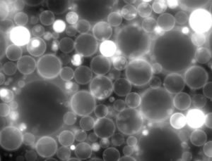
Giant Unilamellar Vesicles (GUVs), and the mammalian COPII proteins, as expressed and purified from insect cell culture. I will utilise cryo-electron microscopy and single-molecule fluorescence in the study of the COPII coat assembly through in vitro reconstitution. My PhD supervisor is Dr Giulia Zanetti, ISMB, Birkbeck.
Laura Pokorny
I studied for an undergraduate degree in Biochemistry at the University of York. In the summer between my second and third year I carried out a 2 month research placement in Paul Pryor’s lab at the Centre for Immunology and Infection at the University of York, where I was identifying chlamydial effector proteins involved in disrupting the trafficking of the bacterium to the host lysosome. I really loved working in a research setting and this was when I realised I wanted to do a PhD and pursue a career in research.
Rotation projects (Year 1)
Rotation 1: My first rotation ‘Manipulation of Nuclear Function by Chlamydia trachomatis’ was in Dr Richard Hayward’s lab (ISMB, Birkbeck). Previous research in the Hayward lab had identified alterations in nuclear architecture during infection by C. trachomatis. Namely, the nuclear shape becomes distorted in infected cells, lamin A/C is decreased at the inclusion distal face of the nucleus, and there was a degradation of nucleoporins at the inclusion proximal face of the nucleus. I confirmed these findings by aiming to understand the mechanism underlying the lamin A/C decrease.

Caspase 6 is a candidate for the degredation of lamin A/C due to the fact that lamin A/C is degraded by caspase 6 during apoptosis. By treating infected cells with a drug which inhibits caspase 6, I was able to block the lamin A/C decrease in infected cells. This was shown by confocal microscopy and by western blot.
Rotation 2: My second rotation ‘A novel mechanism of targeting and transport of a P. falciparum protein down the secretory pathway’ was in Dr Andrew Osborne’s lab (ISMB, UCL). The mechanism leading to protein transport, and in particular trans-membrane protein transport, in P. falciparum is not completely understood. Proteins destined for export must cross many membranes of the parasite before entering the host cell. Models have proposed whereby TM proteins are extracted from membranes at various stages of the secretory pathways and trafficked via chaperones (Papakrivos, Newbold and Lingelbach., 2005; Kneupfer et al., 2005; Gruring et al., 2012). However, the concept of pulling proteins out of membranes during protein export is unsupported outside the Plasmodium field. Recent work in the Osborne lab and others has provided evidence that the PNEP protein Pf332, which has a single TM domain, behaves in line with this extraction model. I used yeast as a model organism and showed that, when Pf332 is expressed in yeast, there is a subset of soluble protein. This suggests that the machinery needed to pull the protein out the membranes is conserved in eukaryotes. In this rotation I used techniques including western blotting, parasite culturing, cloning, and florescence microscopy.
Rotation 3: In my third rotation ‘Single-molecule studies of the molecular mechanisms of the nuclear pore complex during C. trachomatis infection’ in Dr Alan Lowe’s lab (ISMB, UCL) I used super-resolution microscopy to gain images of the nucleoporin degradation seen in my first rotation, and to learn more about the kinetics of importin-beta transport in the nucleus during infection. I used the technique of photoactivated localization microscopy (PALM). In short, PALM imaging uses the principle of stochastically activating, imaging and photobleaching photoswitchable fluorescent proteins in order to temporally separate closely spaced molecules (Betzig et al., 2006). The resolution achieved in PALM imaging is over an order of magnitude higher than the diffraction limit of light. By transfecting infected cells with importin-B (nuclear transport receptor) tagged to a photoswitchable fluorescent protein and imaging by PALM, we could gain a much higher resolution picture of the organisation of the nuclear pores, and could follow the kinetics of transport via single-particle tracking.
‘Working within the ISMB environment has been a great way to find out more about the work of other PhD students and staff through weekly presentations during term time known as Friday Wraps’
PhD project: Studying Vaccinia virus fusion using a minimal model system (Year 2 onwards)
Vaccinia virus (VACV) is the prototypical Poxvirus. Poxviruses enter cells by acid mediated fusion, using the most complicated virus fusion machinery identified. Whilst genetics indicates that poxvirus fusion relies on 12 viral proteins, to date the organisation of this machinery, its mechanism of fusion, its fusion peptide, and the structural and molecular details of poxvirus fusion remain a mystery. Therefore to address this lack in our knowledge, I aim to develop a new minimal model system to study VACV entry and fusion. This system will be amenable to super-resolution imaging studies allowing us an unprecedented view into the biological requirements of viral entry. My PhD supervisor is Dr Jason Mercer LMCB.
